Visual Anatomy & Physiology Lab Manuals, like Sarikas’ editions, offer structured learning, aiding comprehension of core concepts through a visual approach.
These manuals, including the Main Version and Pig Version, complement textbooks like Martini/Ober/Nath, enhancing the lab experience for students.
Pearson publishes updated editions, such as the 2nd edition from July 14, 2021, providing current anatomical and physiological insights.
Purpose of Lab Manuals
Visual Anatomy & Physiology Lab Manuals serve as essential companions, bridging theoretical knowledge with practical application. They guide students through dissections, microscope usage, and observation, reinforcing anatomical structures and physiological processes.
These manuals, like those by Sarikas, provide structured exercises and clear instructions, fostering a deeper understanding of the human body.
They aim to develop critical thinking, problem-solving skills, and laboratory techniques crucial for future healthcare professionals, enhancing the overall learning experience.
Target Audience
Visual Anatomy & Physiology Lab Manuals are primarily designed for students enrolled in introductory anatomy and physiology courses, often within allied health programs.
This includes aspiring nurses, medical technicians, physical therapists, and other healthcare professionals needing a solid foundation in human biology.
The manuals cater to diverse learning styles, utilizing visual aids to support students with varying academic backgrounds. They are suitable for both two-year and four-year college curricula.
The Human Body: An Overview
Visual Anatomy & Physiology Lab Manuals facilitate understanding of the human body’s complex organization, from chemical levels to organ systems, aiding comprehensive study.
Levels of Organization
Visual Anatomy & Physiology Lab Manuals effectively demonstrate the hierarchical levels of organization within the human body. Starting with chemical components – atoms and molecules – these manuals build understanding towards cellular structures, the basic units of life.
Further exploration reveals tissue types, groups of similar cells, followed by organ systems comprised of interacting organs.
Ultimately, the organismal level, the complete human being, is visualized and analyzed, providing a holistic perspective reinforced by practical lab exercises.
Anatomical Terminology
Visual Anatomy & Physiology Lab Manuals prioritize mastering precise anatomical terminology. These manuals utilize standardized language to describe body structures and their relationships, crucial for accurate communication.
Students learn directional terms – superior, inferior, anterior, posterior – alongside regional terms identifying specific body areas.
Understanding prefixes, suffixes, and root words is emphasized, enabling dissection and identification of anatomical features.
The lab component reinforces these terms through practical application, solidifying comprehension for future study.
Body Planes and Cavities
Visual Anatomy & Physiology Lab Manuals clearly illustrate body planes – sagittal, frontal (coronal), and transverse – essential for sectioning and viewing anatomical structures.
These manuals detail major body cavities, including dorsal (cranial and vertebral) and ventral (thoracic, abdominal, and pelvic), explaining organ placement.
Understanding these divisions is vital for interpreting imaging techniques and relating structure to function.
Lab exercises often involve identifying structures within specific cavities, reinforcing spatial relationships and anatomical orientation.

Microscopic Anatomy
Visual Anatomy & Physiology Lab Manuals facilitate the study of tissues – epithelial, connective, muscle, and nervous – using microscopy and detailed illustrations.
These resources aid in cell structure identification and understanding functional relationships at the microscopic level.
Cell Structure and Function
Visual Anatomy & Physiology Lab Manuals guide students through examining cell components using microscopy, a crucial skill for understanding life’s building blocks.
Labs focus on identifying organelles – nuclei, cytoplasm, membranes – and correlating structure with function.
These manuals often include exercises on focusing techniques, like raising the microscope tube slowly for clear image acquisition.
Understanding oil immersion (100x objective) is also emphasized, enabling detailed observation of cellular structures.
The manuals support learning about cellular transport, communication, and metabolic processes.
Epithelial Tissue
Visual Anatomy & Physiology Lab Manuals facilitate the study of epithelial tissues through microscopic observation, categorizing them by shape and layering.
Labs guide students in identifying squamous, cuboidal, and columnar epithelia, alongside simple and stratified arrangements.
These manuals often include exercises on recognizing specialized epithelial features like cilia and microvilli, linking structure to function.
Students learn to differentiate between covering and lining epithelium versus glandular epithelium.
The practical exercises reinforce understanding of tissue roles in protection, secretion, absorption, and filtration.
Connective Tissue
Visual Anatomy & Physiology Lab Manuals aid in identifying diverse connective tissues using microscopy, emphasizing matrix composition and cell types.
Labs focus on recognizing connective tissue proper – loose (areolar, adipose, reticular) and dense (regular, irregular, elastic) – and specialized types.
Students learn to differentiate cartilage (hyaline, elastic, fibrocartilage) and bone tissue, noting structural characteristics.
Blood is also examined as a connective tissue, analyzing its components.
Practical exercises connect tissue structure to functions like support, protection, binding, and transport.
Muscle Tissue
Visual Anatomy & Physiology Lab Manuals guide students in identifying the three muscle tissue types: skeletal, smooth, and cardiac, under a microscope.
Labs emphasize distinguishing features like striations, cell shape, and presence of multiple nuclei in skeletal muscle.
Students learn to recognize smooth muscle’s non-striated appearance and location in organ walls.
Cardiac muscle is identified by its striations, branching fibers, and intercalated discs.
Practical exercises correlate microscopic structure with muscle contraction mechanisms and physiological functions.
Nervous Tissue
Visual Anatomy & Physiology Lab Manuals facilitate the study of nervous tissue components: neurons and neuroglia, using microscopy.
Labs focus on identifying neuron structures – cell body, dendrites, axon – and understanding their functional roles in signal transmission.
Students differentiate between various neuroglial cells (astrocytes, oligodendrocytes, microglia) based on morphology and support functions.
Manuals aid in recognizing nerve fiber types (myelinated vs. unmyelinated) and their impact on conduction velocity.
Practical exercises connect microscopic anatomy to nervous system physiology and signal processing.

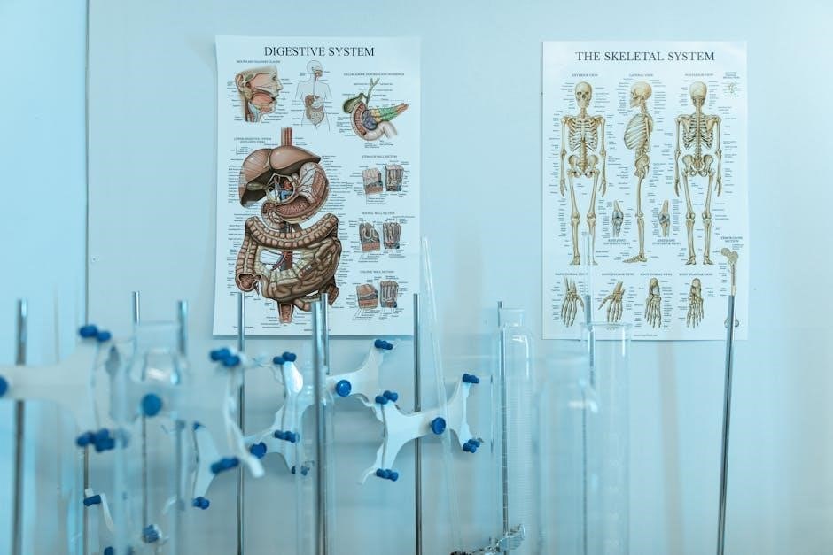
Skeletal System
Visual Anatomy & Physiology Lab Manuals guide bone structure and function studies, classifying bone types and joint mechanics through practical exercises.
Labs emphasize identifying skeletal features and understanding their relationship to movement and support.
Bone Structure and Function
Visual Anatomy & Physiology Lab Manuals facilitate understanding of bone composition, detailing compact and spongy bone tissues.
Labs guide students through identifying bone markings – processes, foramina, and fossae – and correlating them with function.
Practical exercises focus on bone development, remodeling, and the roles of osteoblasts, osteocytes, and osteoclasts;
Manuals emphasize how bone structure supports weight bearing, protection, and mineral storage, linking anatomy to physiological processes.
Students learn to analyze bone histology and relate microscopic features to macroscopic bone shapes.
Types of Bones
Visual Anatomy & Physiology Lab Manuals categorize bones into long, short, flat, irregular, and sesamoid types, using illustrative diagrams.
Labs involve identifying examples of each bone class within the skeletal system, enhancing practical recognition skills.
Students analyze the structural differences between bone types and correlate these with their specific functions.
Manuals detail how bone shape influences movement, support, and protection within the body.
Exercises focus on recognizing bone features unique to each classification, solidifying understanding of skeletal diversity.
Joints
Visual Anatomy & Physiology Lab Manuals detail joint classifications – fibrous, cartilaginous, and synovial – with detailed illustrations.
Labs emphasize identifying joint structures like ligaments, tendons, and cartilage, crucial for stability and movement.
Students explore the range of motion permitted by different joint types, correlating structure with function.
Manuals showcase examples like hinge, ball-and-socket, and pivot joints, aiding practical identification.
Exercises involve analyzing joint pathologies and understanding how structural damage impacts mobility and overall health.

Muscular System
Visual Anatomy & Physiology Lab Manuals illustrate muscle contraction mechanisms and types – skeletal, smooth, and cardiac.
Labs focus on identifying major skeletal muscles and understanding their origins, insertions, and actions.
Students analyze muscle fiber structure and relate it to functional properties.
Muscle Contraction
Visual Anatomy & Physiology Lab Manuals effectively demonstrate the sliding filament theory, detailing actin and myosin interactions crucial for muscle contraction.
Labs often involve microscopic observation of muscle fibers to visualize sarcomeres and understand the role of calcium ions in initiating the contraction process.
Students explore the neuromuscular junction and the transmission of nerve impulses, linking neural control to muscular responses.
Manuals aid in understanding the energy sources for muscle contraction, including ATP and creatine phosphate, and the consequences of oxygen debt.
Practical exercises reinforce the physiological mechanisms behind muscle shortening and relaxation.
Skeletal Muscles
Visual Anatomy & Physiology Lab Manuals facilitate identification of major skeletal muscles through diagrams and dissection exercises, enhancing anatomical understanding.
Labs focus on muscle origins, insertions, actions, and innervation, solidifying knowledge of musculoskeletal function.
Students learn to classify muscles based on their shape and fiber arrangement, relating structure to performance.
Manuals often include activities to assess muscle strength and endurance, connecting theory to practical application.
Understanding leverage and muscle groups working synergistically is reinforced through interactive exercises and case studies.
Smooth and Cardiac Muscle
Visual Anatomy & Physiology Lab Manuals present smooth and cardiac muscle tissues using microscopic images, highlighting key structural differences from skeletal muscle.
Labs emphasize the involuntary nature of these muscle types and their roles in organ systems like the digestive tract and heart.
Students examine cardiac muscle’s intercalated discs and smooth muscle’s spindle shape, relating form to function.
Manuals detail the unique contractile mechanisms and physiological control of each muscle type.
Activities explore the impact of hormones and neurotransmitters on smooth and cardiac muscle activity, deepening understanding;
Nervous System
Visual Anatomy & Physiology Lab Manuals illustrate brain anatomy, the spinal cord, and the peripheral nervous system through diagrams.
Labs focus on identifying structures and understanding neural pathways, enhancing neurological comprehension.
Brain Anatomy
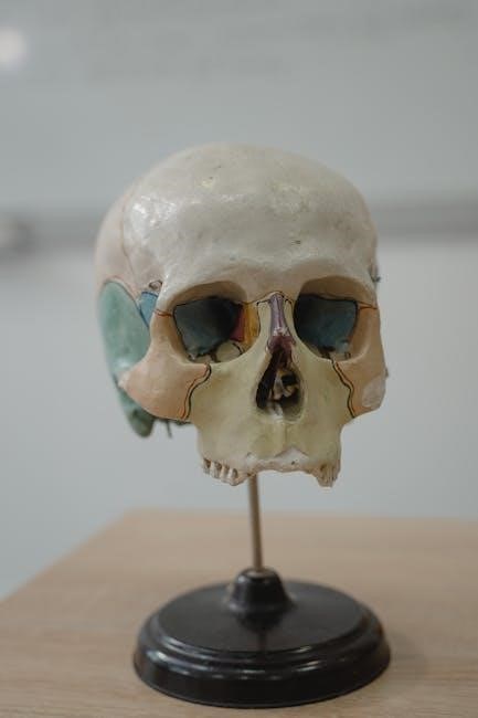
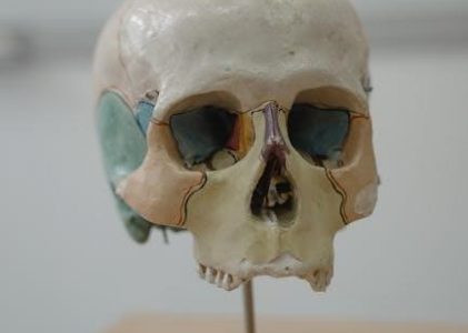
Visual Anatomy & Physiology Lab Manuals dedicate significant sections to detailed brain anatomy exploration. Students utilize these manuals to identify major brain regions, including the cerebrum, cerebellum, and brainstem, through illustrative diagrams and models.
Labs often involve dissecting sheep brains (Pig Version) or utilizing models to pinpoint structures like the frontal, parietal, temporal, and occipital lobes.
Understanding the functions associated with each region is emphasized, alongside tracing neural pathways and recognizing key anatomical landmarks.
These exercises solidify comprehension of complex neurological structures.
Spinal Cord
Visual Anatomy & Physiology Lab Manuals guide students through the intricate anatomy of the spinal cord, emphasizing its protective structures and internal organization. Labs utilize models and diagrams to identify the dorsal and ventral horns, gray and white matter, and spinal nerves.
Students learn to trace nerve pathways and understand the functional significance of different spinal cord segments.
The manuals aid in visualizing the relationship between the spinal cord and peripheral nerves, crucial for understanding reflex arcs and motor control.
Dissection exercises may be included.
Peripheral Nervous System
Visual Anatomy & Physiology Lab Manuals detail the components of the peripheral nervous system, including cranial and spinal nerves, ganglia, and sensory receptors.
Labs focus on identifying nerve distributions and understanding the functional roles of different peripheral nerves, like those controlling muscle movement or sensation.
Students utilize diagrams and models to trace nerve pathways and differentiate between somatic and autonomic nervous systems.
Manuals aid in visualizing the connection between the central and peripheral nervous systems.
Cardiovascular System
Visual Anatomy & Physiology Lab Manuals illustrate heart anatomy, blood vessels, and blood composition.
Labs emphasize tracing blood flow and identifying key structures for understanding cardiovascular function.
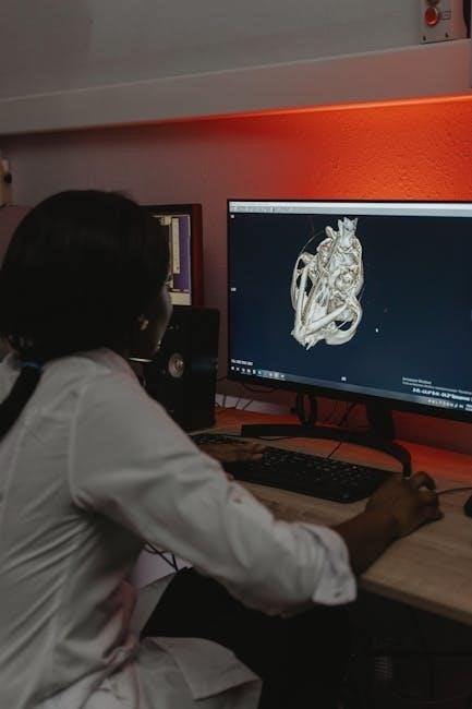
Heart Anatomy
Visual Anatomy & Physiology Lab Manuals provide detailed diagrams and exercises focusing on heart anatomy. Students learn to identify chambers – atria and ventricles – and valves, crucial for understanding blood flow.
Labs often involve dissecting preserved hearts or utilizing models to visualize the coronary arteries, myocardium, and pericardium.
These manuals aid in tracing the path of blood through the heart, from vena cava to pulmonary artery and aorta, reinforcing comprehension of cardiac circulation.
Understanding these structures is fundamental to grasping cardiovascular physiology.
Blood Vessels
Visual Anatomy & Physiology Lab Manuals guide students in identifying arteries, veins, and capillaries, emphasizing structural differences relating to function. Labs often involve tracing vessel pathways using diagrams and models.
Students learn to differentiate between the thick-walled arteries and thinner-walled veins, understanding their roles in pressure regulation and blood return.
Capillaries, the sites of exchange, are also explored, highlighting their microscopic structure.
These manuals reinforce understanding of the circulatory system’s vital role.
Blood Composition
Visual Anatomy & Physiology Lab Manuals facilitate the study of blood components: plasma, red blood cells, white blood cells, and platelets.
Labs often involve microscopic examination of blood smears to identify different cell types and understand their relative proportions.
Students learn to recognize the distinct shapes and features of erythrocytes, leukocytes, and thrombocytes.
Manuals emphasize the functions of each component – oxygen transport, immunity, and clotting – within the circulatory system.
Understanding blood composition is crucial for overall physiological comprehension.
Respiratory System
Visual Anatomy & Physiology Lab Manuals guide dissection and study of lung anatomy, focusing on structures involved in gas exchange;
Labs demonstrate airflow pathways and alveolar function, enhancing understanding of respiration.
Lung Anatomy
Visual Anatomy & Physiology Lab Manuals facilitate detailed exploration of lung anatomy, guiding students through identifying key structures like the trachea, bronchi, and bronchioles.
Labs often involve dissection or model examination to visualize the lobar structure of the lungs, including the right lung’s three lobes and the left lung’s two.
Students learn to differentiate between conducting and respiratory zones, observing the alveoli where gas exchange occurs.
Manuals support understanding of the pleura and its role in lung function, enhancing comprehension of respiratory mechanics.
Gas Exchange
Visual Anatomy & Physiology Lab Manuals illustrate gas exchange processes within the lungs, detailing oxygen and carbon dioxide diffusion across the alveolar-capillary membrane.
Labs often utilize models or diagrams to demonstrate partial pressure gradients driving oxygen into the blood and carbon dioxide out.
Students analyze factors influencing gas exchange efficiency, like surface area and membrane thickness, using manual guidance.
The manuals support understanding of hemoglobin’s role in oxygen transport and the impact of various conditions on respiratory function.
Digestive System
Visual Anatomy & Physiology Lab Manuals detail organ structure, guiding dissection and identification of digestive components.
Labs explore digestive processes, like enzymatic breakdown, using models and simulations for enhanced understanding.
Organ Structure
Visual Anatomy & Physiology Lab Manuals meticulously guide students through the organ structure of the digestive system.
These manuals facilitate identification of key anatomical features within organs like the stomach, small intestine, large intestine, liver, and pancreas.
Dissection exercises, supported by detailed illustrations, allow for hands-on exploration of tissue layers and organ arrangements.
Labs emphasize understanding the relationship between structure and function, clarifying how each organ contributes to the overall digestive process.
Students learn to differentiate between organ components and their roles in nutrient absorption and waste elimination.
Digestive Processes
Visual Anatomy & Physiology Lab Manuals illuminate digestive processes, from ingestion to elimination, with clarity and detail.
Labs explore mechanical digestion – chewing, churning – and chemical digestion, focusing on enzyme action and nutrient breakdown.
Students investigate absorption in the small intestine, tracing the pathways of carbohydrates, proteins, and fats.
Manuals often include exercises on peristalsis and segmentation, demonstrating how these movements propel food through the digestive tract.
Understanding the interplay of organs and enzymes is emphasized, solidifying comprehension of efficient nutrient processing.
Urinary System
Visual Anatomy & Physiology Lab Manuals detail kidney structure and urine formation processes.
Labs explore nephron function, filtration, reabsorption, and secretion, enhancing understanding of renal physiology.
Kidney Structure
Visual Anatomy & Physiology Lab Manuals meticulously illustrate the kidney’s internal and external anatomy. Students explore the renal capsule, cortex, medulla, and the renal pelvis through diagrams and exercises.
Labs focus on identifying nephrons – the functional units – including the glomerulus, Bowman’s capsule, proximal and distal convoluted tubules, and the loop of Henle.
These manuals aid in visualizing blood vessel arrangements, like the afferent and efferent arterioles, crucial for understanding filtration processes within the kidney’s complex structure.
Urine Formation
Visual Anatomy & Physiology Lab Manuals detail the three key processes of urine formation: glomerular filtration, tubular reabsorption, and tubular secretion.
Labs guide students in understanding how blood pressure drives filtration at the glomerulus, creating filtrate. Diagrams illustrate reabsorption of essential substances back into the bloodstream.
Exercises demonstrate how waste products are actively secreted into the tubules, ultimately forming urine. Manuals emphasize the nephron’s role in maintaining fluid and electrolyte balance.
Reproductive System
Visual Anatomy & Physiology Lab Manuals present detailed diagrams of male and female reproductive anatomy, aiding identification of organs.
Labs explore gametogenesis and hormonal control, enhancing understanding of reproductive processes.
Male Reproductive Anatomy
Visual Anatomy & Physiology Lab Manuals meticulously illustrate the male reproductive system, beginning with external structures like the scrotum and penis.
Internal anatomy is detailed, showcasing the testes, epididymis, vas deferens, seminal vesicles, prostate gland, and bulbourethral glands.
Labs often involve identifying these structures on diagrams and models, alongside tracing the pathway of sperm.
The manual aids in understanding the histological features of the testes and associated glands, crucial for hormone production and gametogenesis.
Students learn about the functional relationships between these components.
Female Reproductive Anatomy
Visual Anatomy & Physiology Lab Manuals comprehensively depict the female reproductive system, starting with external genitalia like the vulva.
Internal structures, including the ovaries, fallopian tubes, uterus, cervix, and vagina, are meticulously illustrated for detailed study.
Lab exercises focus on identifying these structures on anatomical models and diagrams, tracing oocyte transport.
Histological examination of the ovary and uterine wall is emphasized, relating structure to hormonal cycles and implantation.
The manual clarifies the functional interplay of these organs during reproduction.

Endocrine System
Visual Anatomy & Physiology Lab Manuals illustrate major endocrine glands – pituitary, thyroid, adrenals, pancreas – and their hormone production.
Labs emphasize hormone regulation mechanisms and effects on target tissues, enhancing understanding of endocrine function.
Major Glands and Hormones
Visual Anatomy & Physiology Lab Manuals meticulously detail major endocrine glands, including the pituitary, thyroid, parathyroid, adrenal, and pancreas.
Labs focus on identifying these glands’ anatomical locations and correlating them with specific hormone production.
Students explore hormones like insulin, glucagon, cortisol, epinephrine, thyroid hormones, and growth hormone.
Manuals often include diagrams and exercises to understand hormone chemical structures and their roles in regulating bodily functions.
Understanding hormone secretion and target cell interactions is a key learning objective.
Hormone Regulation
Visual Anatomy & Physiology Lab Manuals emphasize the intricate feedback loops governing hormone secretion, particularly negative feedback mechanisms.
Labs illustrate how hormone levels influence gland activity, maintaining homeostasis.
Students analyze scenarios demonstrating the hypothalamic-pituitary axis’s control over various endocrine glands.
Manuals often include exercises exploring disruptions in hormone regulation, like those seen in diabetes or thyroid disorders.
Understanding the interplay between hormones and target tissues is crucial, alongside the role of receptors.

Immune System
Visual Anatomy & Physiology Lab Manuals detail innate and adaptive immunity, showcasing immune cell identification.
Labs explore immune responses, emphasizing cellular interactions and antibody function for effective defense.
Innate and Adaptive Immunity
Visual Anatomy & Physiology Lab Manuals effectively demonstrate the distinctions between innate and adaptive immune responses.
Labs often focus on identifying components of innate immunity – physical barriers, cellular defenses like macrophages, and inflammatory responses.
Students explore adaptive immunity through exercises examining lymphocytes (B and T cells), antibody production, and immunological memory.
Manuals utilize visual aids to clarify complex processes like antigen presentation and the activation of immune pathways, solidifying understanding.
Practical exercises may involve simulated immune challenges to observe the body’s defense mechanisms.
Immune Cells
Visual Anatomy & Physiology Lab Manuals provide detailed microscopic views of key immune cells, aiding identification and functional understanding.
Labs commonly feature exercises on recognizing leukocytes – neutrophils, lymphocytes, monocytes, eosinophils, and basophils – based on their morphology.
Students learn to differentiate between B cells, T cells, and macrophages, understanding their roles in adaptive and innate immunity.
Manuals often include diagrams illustrating cell signaling pathways and interactions during immune responses.
Practical activities may involve staining and observing immune cells in blood smears or tissue samples.

Lab Safety and Procedures
Visual Anatomy & Physiology Lab Manuals emphasize safety, covering microscope usage and dissection techniques.
Proper handling of specimens, staining procedures, and waste disposal are crucial components of lab protocols.
Microscope Usage
Visual Anatomy & Physiology Lab Manuals detail proper microscope operation for observing microscopic structures. Begin with the lowest power objective, gradually increasing magnification.
Carefully raise the tube while focusing, ensuring a clear image. For oil immersion (100x), apply a drop of immersion oil to the slide before engaging the objective.
Clean lenses only with lens paper. Proper illumination adjustment is vital for optimal viewing. Always carry the microscope with both hands – one on the arm, the other under the base.
Dissection Techniques
Visual Anatomy & Physiology Lab Manuals emphasize careful dissection for understanding anatomical relationships. Utilize appropriate dissection tools – scalpels, scissors, and probes – with precision and respect;
Follow the manual’s instructions meticulously, identifying structures as you expose them. Proper pinning techniques secure tissues for clear visualization.
Record observations accurately through drawings or photographs. Dispose of biological waste responsibly, adhering to lab safety protocols. Gentle handling minimizes tissue damage, enhancing learning.