Article Plan: Meniscus Tear Exercises PDF
This comprehensive guide details a phased rehabilitation approach, incorporating exercises for range of motion, strengthening, and proprioception.
It addresses both conservative and post-operative management,
with a focus on avoiding meniscal loading during recovery, as per current standards of care.
Meniscal tears are common knee injuries, often occurring during sports or from degenerative changes. Understanding these injuries is crucial for effective rehabilitation. This article provides a detailed plan for meniscus tear exercises, encompassing conservative management and post-operative protocols. The information presented aims to guide patients and clinicians through a structured recovery process, optimizing functional outcomes and minimizing the risk of re-injury.
Effective rehabilitation, as highlighted in recent research, focuses on restoring knee function through targeted exercises. These exercises address pain, swelling, and limitations in range of motion. A systematic approach, incorporating progressive loading and proprioceptive training, is essential. Conservative management, often the initial approach, utilizes physical therapy to strengthen surrounding muscles and improve stability. However, surgical intervention may be necessary for certain tear types or when conservative measures fail. This guide will cover both pathways, providing a comprehensive resource for meniscus tear rehabilitation.
The protocols discussed are based on current best practices and aim to facilitate a safe and efficient return to activity.
Understanding the Meniscus
The menisci are C-shaped fibrocartilage structures within the knee joint, vital for shock absorption and load distribution. They contribute significantly to knee stability and allow for smooth articulation between the femur and tibia. Comprehending their function is paramount when designing effective exercise programs following a tear. Each knee possesses two menisci – medial and lateral – and their integrity is crucial for optimal knee biomechanics.
These structures aren’t simply passive cushions; they actively participate in joint proprioception, providing feedback about joint position and movement. Damage to the meniscus disrupts this feedback, potentially leading to instability and increased risk of further injury. Rehabilitation protocols, therefore, must address proprioceptive deficits alongside strength and range of motion. Understanding the specific type of tear – vertical, horizontal, or bucket-handle – is also essential, as it influences the rehabilitation timeline and exercise selection.
Properly understanding the meniscus allows for targeted rehabilitation.
Function of the Meniscus
The meniscus performs several critical functions within the knee joint. Primarily, it deepens the articular surfaces of the tibia and femur, enhancing joint congruity and stability. This improved fit increases the contact area, distributing weight-bearing forces more evenly and reducing stress on the articular cartilage – preventing premature osteoarthritis.
Beyond load transmission, the menisci act as shock absorbers, attenuating compressive forces during activities like walking, running, and jumping. They contribute approximately 20-25% to axial load absorption. Furthermore, they play a vital role in joint lubrication, facilitating smooth, frictionless movement.
The menisci also contribute to joint proprioception, providing sensory feedback regarding joint position and movement. This feedback is crucial for maintaining balance and coordinating muscle activity. Damage to the meniscus compromises these functions, leading to pain, instability, and an increased risk of further knee injury. Rehabilitation aims to restore as much of this functionality as possible.
Types of Meniscal Tears
Meniscal tears present in various patterns, influencing rehabilitation approaches. Vertical tears, running along the length of the meniscus, are common and can displace, causing locking or catching sensations. Horizontal tears, extending across the width, often result from shear forces and may cause pain and swelling.
Bucket-handle tears, a severe form of vertical tear, involve a large fragment detaching and displacing into the intercondylar notch, frequently causing mechanical symptoms. Radial tears originate from the inner edge and are often degenerative. Complex or combination tears involve multiple tear patterns.
Tear location – medial or lateral meniscus – also impacts treatment. The medial meniscus is more frequently injured due to its greater attachment to the collateral ligament. The specific tear type dictates the extent of surgical intervention, if required, and influences the progression of rehabilitation exercises; Accurate diagnosis is crucial for tailoring an effective recovery plan.
Conservative Management of Meniscus Tears
Not all meniscus tears require surgery; conservative management is often the initial approach. This focuses on reducing pain, swelling, and restoring function through non-operative methods. It’s particularly suitable for stable tears without mechanical symptoms like locking. The Brigham and Women’s Hospital highlights conservative care as a standard approach.
Initial treatment typically involves the RICE protocol – Rest, Ice, Compression, and Elevation – to manage acute inflammation. Pain medication, such as over-the-counter analgesics, can provide symptomatic relief. Crucially, physical therapy plays a central role, focusing on strengthening surrounding muscles to support the knee joint and improve stability.
The goal is to enhance proprioception, the body’s awareness of its position in space, and restore a full range of motion. Conservative management aims to allow the body to heal naturally, avoiding the risks associated with surgical intervention when appropriate.
When to Consider Non-Surgical Treatment
Non-surgical treatment is often the preferred first line of defense for many meniscus tears, particularly those that are stable and don’t cause mechanical symptoms like locking or catching. Factors influencing this decision include the tear’s size, location, and the patient’s activity level and overall health.
If pain is manageable with conservative measures and the knee remains functional, surgery may be delayed or avoided altogether. Hegedus et al.’s systematic review emphasizes the importance of accurate physical examination to guide treatment decisions. A trial of physical therapy, typically lasting several weeks, is crucial to assess the potential for improvement without surgical intervention.
Patients with degenerative tears, common in older adults, often respond well to non-surgical management. The focus shifts to symptom relief and maintaining function rather than repairing the tear itself. Ultimately, the decision is made collaboratively between the patient and healthcare provider, considering individual circumstances.
Role of Physical Therapy
Physical therapy is central to both non-surgical and post-operative meniscus tear rehabilitation. Its role extends beyond simply prescribing exercises; it involves a comprehensive assessment to identify impairments in strength, range of motion, proprioception, and functional movement patterns.
A skilled physical therapist will design a tailored program progressing through phases, starting with pain and swelling management, followed by restoring knee motion and activating key muscle groups. Strengthening exercises, particularly for the quadriceps and hamstrings, are crucial for stabilizing the knee joint.
Proprioceptive training, focusing on balance and coordination, helps regain neuromuscular control. The therapist will also educate the patient on activity modification and proper biomechanics to prevent re-injury. As highlighted in rehabilitation protocols, avoiding meniscal loading during early stages is paramount, often achieved through open-chain exercises.
Phases of Rehabilitation
Rehabilitation following a meniscus tear is typically structured into three overlapping phases: acute, intermediate, and advanced; Each phase builds upon the previous one, progressively increasing the demands placed on the knee joint.

Phase 1 (0-4 weeks) focuses on controlling pain and inflammation, restoring initial range of motion, and activating key muscles without stressing the meniscus. Bracing, as seen in post-transplant protocols, may be utilized initially.
Phase 2 (4-8 weeks) emphasizes strengthening the surrounding musculature – quadriceps, hamstrings, and calves – alongside proprioceptive exercises to improve balance and coordination. Weight-bearing is gradually increased.
Phase 3 (8+ weeks) concentrates on returning to functional activities and sport-specific training. This phase involves more challenging exercises designed to simulate real-life movements, preparing the patient for full activity. Progression is guided by pain levels and functional milestones.

Phase 1: Acute Phase (0-4 Weeks)
The initial acute phase, lasting approximately 0-4 weeks, prioritizes reducing pain, swelling, and protecting the injured meniscus. A knee brace, potentially locked in extension as per some post-operative protocols, may be employed for immobilization and support during this period.
Goals of Phase 1 include regaining full knee extension, achieving at least 90 degrees of flexion (though flexion beyond 90º may be limited for up to 8 weeks in some repairs), and initiating gentle muscle activation. Avoiding meniscal loading is crucial.
Exercises focus on pain-free range of motion exercises like heel slides and gentle hamstring stretches. Isometric quadriceps sets and gluteal squeezes help activate muscles without joint movement. Ankle pumps promote circulation. Emphasis is on low-impact activities and avoiding any movements that exacerbate symptoms.
Goals of Phase 1
The primary goals during the initial 0-4 week acute phase are centered around mitigating immediate symptoms and establishing a foundation for subsequent rehabilitation. Reducing pain and swelling are paramount, achieved through controlled movement and minimizing stress on the injured meniscus.
Specifically, we aim to restore full passive knee extension, which is vital for normal gait. Achieving at least 90 degrees of knee flexion is a key milestone, though protocols may limit this initially, particularly post-surgery. Initiating voluntary muscle activation, specifically of the quadriceps and gluteals, is crucial to prevent atrophy.
Furthermore, protecting the healing meniscus is essential, avoiding any activities that could aggravate the tear or compromise repair. Establishing proper neuromuscular control and preparing the patient for the progression to more demanding exercises in Phase 2 are also key objectives.
Exercises for Phase 1: Range of Motion & Muscle Activation

Initial exercises focus on gentle, pain-free movements. Ankle pumps promote circulation and reduce swelling. Heel slides, performed lying down, gradually increase knee flexion, aiming for that 90-degree target, respecting any bracing limitations. Quadriceps sets – contracting the thigh muscle without moving the knee – initiate muscle activation without joint loading.
Short-arc quads (extending the knee slightly from a bent position) further strengthen the quadriceps. Straight leg raises, performed with the knee locked, engage the quads and hip flexors. Gentle hamstring sets help maintain muscle balance.
Proprioceptive awareness can be started with weight shifts while seated, encouraging subtle muscle engagement. Avoid any twisting or pivoting motions. Crucially, all exercises should be performed within a pain-free range, and a knee brace may be utilized as prescribed, particularly in the early stages post-operatively, as described in rehabilitation protocols.
Phase 2: Intermediate Phase (4-8 Weeks)
Phase 2 builds upon the foundation of Phase 1, progressively increasing load and complexity. Strengthening exercises become more prominent, focusing on controlled movements. Wall squats, initiated with a limited range of motion, enhance quadriceps and gluteal strength. Step-ups onto a low platform improve functional strength and stability.
Hamstring curls (using resistance bands or a machine) target the posterior chain. Calf raises address lower leg strength. Proprioceptive exercises advance to include single-leg stance with minimal wobble, and balance board activities, challenging stability.
Closed kinetic chain exercises, like mini-lunges, are introduced cautiously, avoiding deep flexion to minimize meniscal stress. The focus remains on maintaining proper form and pain-free movement. Post-operative protocols often emphasize open kinetic chain quadriceps work to avoid loading the repair, particularly during the initial 8 weeks, as detailed in meniscal repair guidelines.
Goals of Phase 2
The primary goals of Phase 2 (weeks 4-8) center around restoring strength and improving neuromuscular control. This phase transitions from pain management and initial range of motion to actively rebuilding muscle support around the knee joint. Increasing quadriceps strength is crucial, but must be achieved without exacerbating meniscal irritation.
Proprioception, or the sense of joint position, is a key focus, aiming to regain stability and prevent re-injury. Improving functional movement patterns, such as walking and stair climbing, is also essential. The aim is to gradually increase weight-bearing tolerance and prepare the knee for more demanding activities.
Reducing swelling and maintaining a pain-free range of motion remain important considerations. Progress is guided by symptom response, with modifications made as needed. Ultimately, Phase 2 aims to establish a solid foundation for the advanced rehabilitation phase, enabling a return to higher-level function.
Exercises for Phase 2: Strengthening & Proprioception
Strengthening exercises in Phase 2 focus on the quadriceps, hamstrings, and calf muscles. Open kinetic chain exercises, like leg extensions and hamstring curls, are favored to minimize direct meniscal loading. Progressive resistance is added as tolerated. Short arc quads and straight leg raises continue to build foundational strength.
Proprioceptive training utilizes balance exercises, such as single-leg stance (with support initially), wobble board activities, and perturbation training. These drills challenge the knee’s ability to maintain stability and react to unexpected movements.
Functional exercises, like step-ups and mini-squats, are introduced cautiously, ensuring proper form and avoiding deep flexion beyond 90 degrees for up to 8 weeks post-injury or surgery. The focus remains on controlled movements and avoiding pain. Core strengthening is also incorporated to enhance overall stability and movement control.
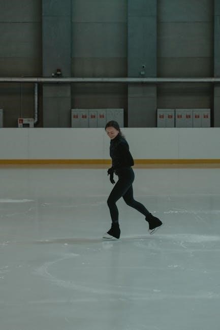
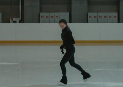
Phase 3: Advanced Phase (8+ Weeks)
Phase 3 concentrates on a return to activity and functional training, gradually increasing the intensity and complexity of exercises. Plyometrics, including jumping and hopping drills, are introduced to improve power and agility, but only when pain-free and with excellent control.
Sport-specific drills are incorporated to simulate the demands of the individual’s chosen activity. This may involve running, cutting, pivoting, and agility exercises. Progressive loading is crucial, carefully monitoring for any signs of re-injury or increased pain.
Continued strengthening and proprioceptive exercises maintain the gains achieved in previous phases. Emphasis is placed on eccentric strengthening to enhance muscle control during deceleration. A successful return to activity requires meeting specific functional criteria, including full range of motion, adequate strength, and good proprioception.
Goals of Phase 3
The primary goals of Phase 3 are to restore full functional capacity and facilitate a safe return to pre-injury activity levels. This involves maximizing strength, power, endurance, and agility, while refining neuromuscular control and proprioception.
Patients should achieve near-symmetrical limb symmetry in strength and functional testing; The focus shifts from simply reducing pain and inflammation to optimizing performance and preventing re-injury.
A key objective is to regain confidence in the knee’s stability and ability to withstand the demands of daily life and sport. This phase aims to prepare the individual for the physical stresses encountered during activity, ensuring they can participate fully and without apprehension. Ultimately, the goal is a sustainable return to activity with minimal risk of recurrence.

Exercises for Phase 3: Return to Activity & Functional Training
This phase incorporates advanced plyometrics, including box jumps, lateral hops, and depth jumps, to enhance power and explosiveness. Agility drills, such as shuttle runs and cone drills, improve quickness and change-of-direction ability.
Sport-specific training is crucial, mimicking the movements and demands of the individual’s chosen activity. This may involve running, cutting, pivoting, and jumping exercises tailored to their sport. Progressive loading is key, gradually increasing the intensity and volume of training.
Functional exercises like lunges with rotation, single-leg squats, and step-ups with weight challenge stability and strength in dynamic movements. A return-to-sport progression should be followed, ensuring criteria are met at each stage before advancing. Monitoring for pain or swelling is vital throughout this phase.
Specific Exercises for Meniscus Tear Rehabilitation
Quadriceps exercises include straight leg raises, wall sits, and leg extensions (with caution, avoiding full extension initially). Hamstring curls, bridge exercises, and Nordic hamstring curls strengthen the posterior chain, providing knee stability.
Calf raises (both straight-knee and bent-knee) improve ankle and lower leg strength, contributing to overall lower limb function. Proprioceptive exercises are vital for regaining balance and coordination.
These include single-leg stance, wobble board exercises, and balance beam walks. Closed kinetic chain exercises, like squats and lunges, are favored to minimize stress on the meniscus. Open kinetic chain exercises should be introduced cautiously, focusing on controlled movements. Progressive resistance is applied as strength improves, always monitoring for pain.
Quadriceps Strengthening Exercises
Quadriceps strength is crucial for knee stability and function post-meniscus injury. Straight Leg Raises (SLR) are foundational, performed lying supine, focusing on controlled hip and knee extension. Wall sits build isometric strength, maintaining a 45-90 degree knee bend.
Leg extensions should be approached cautiously, avoiding full extension to minimize meniscal stress, particularly in early phases. Step-ups onto a low platform progressively increase load. Lunges (forward, reverse, lateral) enhance functional strength and balance.
Focus on open kinetic chain exercises with controlled movements, avoiding forceful contractions. Resistance bands or ankle weights can be added as strength improves. Prioritize proper form to prevent re-injury. Post-operative protocols often emphasize quadriceps strengthening in the open kinetic chain to avoid loading the meniscus;
Hamstring Strengthening Exercises
Hamstring strength balances the quadriceps, contributing to knee stability and preventing re-injury. Hamstring curls, performed with resistance bands or machines, isolate the muscle group. Glute-ham raises are advanced, requiring significant strength and control.
Bridging exercises engage the hamstrings and glutes simultaneously, promoting hip extension and core stability. Romanian Deadlifts (RDLs) with light weights improve hamstring flexibility and strength. Good Mornings, performed with caution, further challenge the posterior chain.
Focus on controlled eccentric contractions, lengthening the hamstring under load. Avoid hyperextension of the knee. Incorporate exercises that mimic functional movements, like walking and running. Strengthening the hamstrings is vital for overall knee health and rehabilitation, complementing quadriceps work.
Calf Strengthening Exercises
Calf muscles contribute to ankle and knee stability, impacting overall lower limb function. Calf raises, both seated and standing, target the gastrocnemius and soleus muscles. Variations include performing them with a slight knee bend to emphasize the soleus. Resistance bands can increase the challenge.

Single-leg calf raises enhance balance and proprioception, crucial for functional recovery. Plyometric calf exercises, like jump rope or box jumps (progressed cautiously), improve power and explosiveness. Eccentric calf raises, slowly lowering the heel, strengthen the muscle during lengthening.
Proper form is essential – maintain a controlled range of motion and avoid bouncing. Strengthening the calves supports the knee joint and improves gait mechanics. Integrate calf exercises into a comprehensive rehabilitation program for optimal results, complementing hamstring and quadriceps work.
Proprioceptive Exercises

Proprioception, or body awareness, is vital for knee stability and preventing re-injury. Single-leg stance, starting with eyes open and progressing to eyes closed, challenges balance. Wobble board or balance disc exercises further enhance stability, requiring constant adjustments.
Perturbation training, where a therapist gently pushes or pulls the patient during stance, improves reactive neuromuscular control. Agility drills, like cone drills and shuttle runs (progressed gradually), integrate proprioception into dynamic movements. Star excursion balance test objectively measures dynamic balance and identifies deficits.
Focus on maintaining proper alignment and controlled movements. Proprioceptive exercises retrain the nervous system to respond effectively to changes in joint position. These drills are crucial in the intermediate and advanced phases of rehabilitation, preparing the knee for return to activity and functional tasks.
Post-Operative Rehabilitation Protocol
Following arthroscopic meniscus repair, a structured protocol is essential. Initial bracing and weight-bearing restrictions, often involving a locked knee brace for the first four weeks, protect the repair. Progressive weight-bearing is introduced based on surgeon’s guidelines and pain levels.
Early focus is on quadriceps activation in open kinetic chain exercises to minimize meniscal loading. Flexion beyond 90 degrees is typically limited for eight weeks to prevent stress on the repair. Range of motion exercises are carefully progressed, avoiding twisting or pivoting motions.

Rehabilitation emphasizes avoiding meniscal loading during the initial phases. Closed kinetic chain exercises are introduced gradually as healing progresses. Full return to activity is dependent on achieving specific functional milestones, including strength, stability, and pain-free movement, guided by a physical therapist.
Bracing and Weight-Bearing Restrictions
Post-operative bracing is a cornerstone of meniscus repair rehabilitation. A locked knee brace, commonly utilized in Phase one, provides stability and protects the surgical site. Progressive loading is permitted, gradually increasing weight-bearing tolerance over the initial four to six weeks.
Weight-bearing restrictions vary based on the tear’s complexity and surgical technique. Partial weight-bearing, using crutches, is often prescribed initially, progressing to full weight-bearing as tolerated. Brace adjustments are made by the surgeon or physical therapist, monitoring for pain and swelling.
The duration of bracing depends on the repair’s nature; some protocols extend bracing beyond eight weeks. Adherence to weight-bearing guidelines is crucial to prevent re-tear or delayed healing. Regular monitoring ensures appropriate progression and minimizes complications, optimizing recovery outcomes.
Avoiding Meniscal Loading During Rehab
Protecting the repaired meniscus is paramount during rehabilitation. Quadriceps strengthening is often performed in an open kinetic chain position, minimizing direct stress on the healing tissue. This technique isolates the quadriceps without compressing the meniscus.
Exercises that involve deep knee flexion, twisting motions, or high-impact activities are initially avoided. Focus shifts to controlled movements, gradually increasing range of motion while maintaining proper form. Proprioceptive exercises enhance joint stability, reducing the risk of re-injury.
Monitoring for pain and swelling is essential; any increase signals excessive loading. Rehabilitation protocols emphasize a gradual progression, respecting tissue healing times. Avoiding meniscal loading ensures optimal repair integration and a successful return to function, guided by a physical therapist’s expertise.
Resources & PDF Downloads
Accessing reliable information is crucial for successful meniscus tear rehabilitation. The Brigham and Women’s Hospital offers resources on conservative management, outlining non-surgical approaches. PMC (PubMed Central) provides access to research articles detailing rehabilitation protocols post-meniscus transplantation and repair.
Texas Health Sports Medicine provides downloadable rehabilitation guidelines for arthroscopic meniscus repairs, covering vertical, horizontal, and bucket-handle tears. These PDFs detail phased exercises, bracing protocols, and weight-bearing restrictions.
Further research on physical examination tests for assessing meniscal tears can be found through systematic reviews. Consulting with a physical therapist is recommended for personalized exercise programs. Downloadable PDFs offer a convenient way to track progress and adhere to a structured rehabilitation plan, ensuring optimal recovery.